What are the Different Parts of a Tooth?

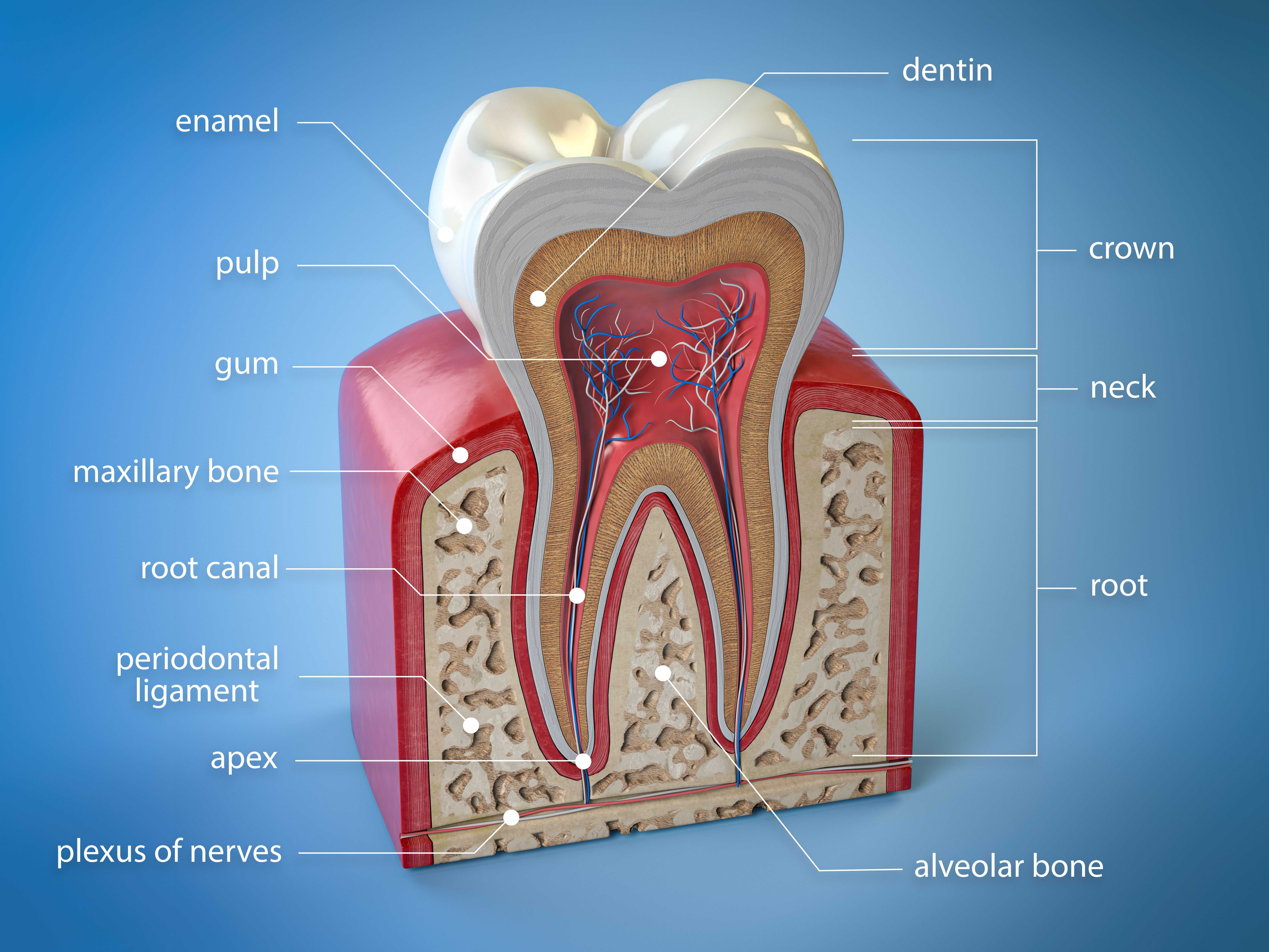
A typical adult has 32 teeth; kids have 20. Even though our teeth are shaped differently, they’re each made up of the same anatomical pieces and layers. The better you understand the different parts of a tooth, the easier it will be for you to understand what your dental provider is talking about during your next checkup!
The Basic Parts of a Tooth
1. The Crown
Most people think of a “cap” that covers their teeth whenever they hear the word “crown.” But a crown is also an important anatomical part of your tooth.
The crown is the white portion of your tooth that extends above the gumlines. It’s covered with enamel. Depending on which tooth it is, your crown may have different shapes. Some tooth crowns are best for biting into and cutting foods, while others serve to grind food up more effectively for digestion.
The back teeth, like molars and premolars, have pointed “cusps” on top of them, with narrow grooves and fissures between them across the chewing surfaces. If you have a young child, your dentist will probably recommend placing sealants on the grooves since those spaces can easily trap food debris and be more prone to cavities.
2. The Root
Your tooth root starts where your crown tapers off, close to the gumlines. Some teeth have one root; others have two or three. Most of the time, they’re fairly straight or have a slight curve to them. And sometimes they’ll even be bent over like a hook (which can make extracting them a bit challenging.)
The tip of each tooth root is called the “apex.” There’s an opening at the apex for the blood and nerve vessels to pass through.
3. Tooth Enamel
Enamel covers your anatomical crown. It tapers off right along the gumlines.
Tooth enamel is the hardest substance in the entire human body, including bones. But if you do wear through or chip your tooth enamel, you’ll likely see the dentin layer underneath.
Any time your dentist spots a cavity in your tooth enamel, they will want to address it before it ruptures into the next layer (dentin,) which isn’t as strong.
4. Dentin
Dentin makes up the bulk of your tooth. It’s what’s immediately underneath your enamel and makes up nearly your entire tooth root.
Dentin isn’t nearly as dense as tooth enamel is, so we always have to worry about our oral health anytime it’s exposed. Acids, decay, and bacteria can eat away at your dentin more quickly than it does your harder tooth enamel. When dentin comes into contact with physical stimuli—whether it’s foods, a toothbrush, cold air, or something else—it tends to be a lot more sensitive than the parts of your tooth that are covered in enamel.
Dentin is also more yellow or brown than enamel, which can affect the color of your smile overall. But that’s not necessarily a bad thing! Since baby (primary) teeth have far less dentin and more enamel than adult teeth, those teeth typically look much whiter than permanent teeth do. You’ll likely notice the difference in color once your child’s adult tooth starts growing right next to one of their primary teeth.
5. Nerve or “Pulp”
Every tooth is a living entity, and as such, it has a nerve and blood supply that feeds it. The tooth nerve runs from the middle of the crown down through the root(s) of the tooth.
If the nerve of your tooth is infected, it usually causes an abscess somewhere near the apex of your tooth root. The swelling and fluid that builds up can drain through the bottom of the root and out through the bone and gum tissue next to it.
During a root canal treatment, your dentist removes the nerve tissues, cleans out the canal that’s left behind, and seals it off. This prevents the inside of your tooth from becoming re-infected. But at that point, it also means your tooth is no longer alive, so you’ll probably need a restorative crown on top of it to protect it.
6. Cementum
Cementum is a very, very thin layer that covers the outside surface of the dentin on your tooth roots. You really can’t even see it without a microscope. The cementum provides a surface for your PDL (mentioned below) to attach.
7. Periodontal Ligament (PDL)
Your tooth root is covered with thousands of microscopic, stretchy little ligaments. These ligaments are what attach the tooth root to the gums and bone around it. They can stretch and pull or even get bruised. If you get hit in the mouth, your PDL may be sore for a few days and need some time to “tighten back up” or spring back from your injury.
8. The “CEJ”
The “cementoenamel junction” or “CEJ” is the neck of your tooth. It’s where the cementum layer over the outside of the root meets the enamel of your crown. Typically, we want to see the CEJ covered by the gum tissues just slightly. If your gums recede just a little bit, you can see a visible margin where the crown tapers off.
9. Alveolar Bone
Ok, so this one isn’t technically a part of the tooth, but it is the anatomical structure immediately around the root of your tooth that helps hold it in place. If you have active gum disease with “pocketing” around your tooth root, there’s a good chance the bone in those spaces is shrinking as well. The less bone you have, the more likely you are to lose your tooth.
Why You Need to Know the Parts of a Tooth
Now that you’re familiar with all of the parts of a tooth, it’s easy to see why your dentist needs to take X-rays during your checkup. If they only looked at your teeth with a light and mirror, they would never see past the crown or enamel. X-rays allow your dentist to see between teeth, around the roots, inside of the dentin, and even assess their bone support. Just because you can’t see it, doesn’t mean your dentist doesn’t need to examine it during your checkup!
Make your inbox smile!
Subscribe
